X-ray Crystallography
X-ray crystallography is a powerful analytical technique used to determine the three-dimensional atomic structure of crystalline materials. It is commonly referred to as single-crystal X-ray diffraction (SCXRD) and is one of the most important tools in materials science, chemistry, biology, and physics for studying the arrangement of atoms within a crystal. This technique allows for precise determination of crystal structures, making it essential for characterizing molecules, minerals, metals, and complex biological macromolecules like proteins and DNA.
Fundamentals of X-ray Crystallography
X-ray crystallography works by directing X-rays at a single crystal, which then diffract as they pass through the ordered arrangement of atoms within the crystal lattice. The resulting diffraction pattern provides information about the atomic arrangement in the crystal, which can be interpreted to reveal the structure of the material at the atomic or molecular level.
The technique is based on Bragg’s law, which relates the diffraction angles to the spacing between atomic planes in the crystal:
nλ=2d sin θ
where:
- n is the diffraction order (an integer),
- λ is the wavelength of the X-rays,
- d is the distance between planes of atoms in the crystal (interplanar spacing),
- θ is half the scattering angle 2θ
Workflow of X-ray Crystallography
The process of single-crystal X-ray diffraction involves several key steps:
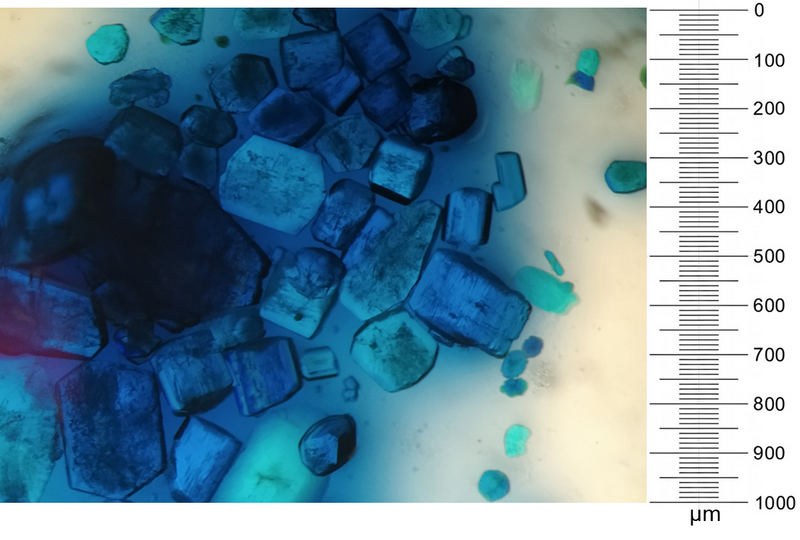
- Crystal preparation or growth: The sample has to be a single, high-quality crystal and can be taken from a natural source or grown synthetically. A good sample is crucial because the quality of the diffraction data depends on the crystal’s ability to diffract X-rays uniformly. Figure 2 shows an example of a batch of freshly grown single crystals of which most would be suitable for single-crystal diffraction.
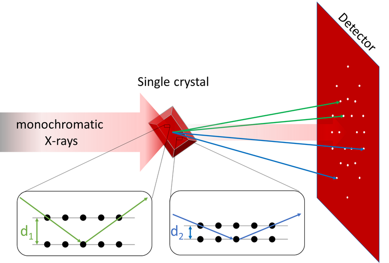
- X-ray exposure: The crystal is placed in an X-ray beam, and as X-rays pass through the crystal, they are diffracted by the regularly arranged atoms within the crystal lattice.
- Data collection: The diffracted X-rays create a pattern of spots on a detector. This pattern, known as a diffraction pattern, is a visual representation of how the crystal structure scatters the X-rays (see Figure 3).
- Data analysis: The intensities and positions of the diffraction spots are recorded and used to calculate the electron density within the crystal. Advanced computational techniques are then applied to reconstruct the positions of the atoms in the crystal.
- Structure refinement: The atomic positions are refined using least square refinement techniques to create a detailed and precise model of the crystal structure, providing insights into bond lengths, angles, and (molecular) geometry.
Crystal Structure Determination
X-ray crystallography is the most precise method for determining the three-dimensional arrangement of atoms in a crystal. The key information obtained from X-ray crystallography includes:
- Unit cell dimensions: The unit cell is the smallest repeating unit in a crystalline material. X-ray diffraction provides the exact dimensions of the unit cell, which is essential for understanding the overall crystal lattice.
- Symmetry and space group: The symmetry of the crystal structure is determined, including its space group, which describes how the atoms are arranged and repeated throughout the crystal. The space group is vital for classifying the crystal structure and for solving complex structures
- Atom positions: The diffraction data is used to calculate the positions of atoms within the unit cell, which helps in determining bond lengths and angles between atoms.
- Types of atoms: Using the electron density map allows not only the determination of atomic positions, but also makes it possible to identify which atoms occupy the position.
Applications of X-ray Crystallography
X-ray crystallography has wide-ranging applications across various scientific disciplines:
Chemistry
In chemistry, X-ray crystallography is used to determine the molecular structures of organic and inorganic compounds. This information is crucial for understanding the reactivity, stability, and properties of chemical compounds. Crystallography is also used to identify polymorphs, which are different crystal structures of the same chemical compound.
Materials science
In materials science, X-ray crystallography helps in analyzing the structure of metals, alloys, ceramics, and minerals. The technique is essential for developing new materials with tailored properties, such as strength, conductivity, and magnetism.
Biology
X-ray crystallography is an indispensable tool in structural biology for determining the structure of biological macromolecules like proteins, enzymes, and nucleic acids (e.g., DNA). The ability to visualize the atomic structure of these molecules helps researchers understand their function and design targeted drugs. The famous discovery of the DNA double helix by Watson and Crick was made possible using X-ray diffraction data.
Pharmaceuticals
Pharmaceutical industry uses X-ray crystallography to study the crystal structures of active pharmaceutical ingredients (APIs). This is crucial for drug design, formulation, and ensuring proper stability and efficacy of the drug.
Geology and mineralogy
Geologists and mineralogists use X-ray crystallography to identify and study the structures of minerals. This information is essential for understanding the formation processes of rocks and minerals and their chemical and physical properties.
Advantages of X-ray Crystallography
- Atomic-scale resolution: X-ray crystallography provides detailed information about the positions of atoms in a crystal, offering an atomic-scale view of the material.
- Accurate structure determination: The technique is highly precise and allows for the accurate determination of bond lengths, angles, and (molecular) geometry.
- Wide range of applications: X-ray crystallography is applicable to a variety of materials, including small molecules, proteins, and complex inorganic structures.
- Non-destructive: The method is non-destructive, meaning the crystal remains intact during analysis.
Limitations of X-ray Crystallography
- Crystal quality: The biggest limitation of X-ray crystallography is that it requires high-quality single crystals, which can be difficult to grow for certain compounds or large biological molecules.
- Time-consuming: Growing crystals and collecting data can be a time-consuming process, particularly for large and complex structures.
- Amorphous materials: X-ray crystallography is not suitable for amorphous materials that lack a well-defined crystal lattice.
Conclusion
X-ray crystallography is a vital technique for determining the crystal structures of materials at atomic resolution. Its ability to provide precise and detailed information about molecular geometry, bonding, and elemental composition makes it indispensable in various scientific fields, from chemistry and biology to materials science and geology. Despite its limitations, such as the need for high-quality crystals, X-ray crystallography remains the gold standard for structure determination.
References
- Glusker, J. P., & Trueblood, K. N. (2010). Crystal Structure Analysis: A Primer. Oxford University Press.
- Giacovazzo, C., Monaco, H. L., & Viterbo, D. (1992). Fundamentals of Crystallography. Oxford University Press.
- Rhodes, G. (2006). Crystallography Made Crystal Clear: A Guide for Users of Macromolecular Models. Academic Press.