mRNA/Liposome-based vaccines
Lots of vaccines are formulated within liposomes. The lipid-based liposome carrier helps protect the mRNA from RNA-cleaving enzymes. These carriers also help bring the mRNA molecules into the target cells. All of this makes consistent liposome size an essential step in vaccine development. One way to monitor liposome particle size and particle size distribution is dynamic light scattering (DLS). Electrophoretic light scattering (ELS) is a useful technique to ensure the stability of the formulation. For the characterization of sub-nanometer dynamics within liposomes, small-angle X-ray scattering (SAXS) is a useful tool.
Read more below about our solutions for tracking and optimizing liposome size and stability, and how to characterize internal lamellar structures, lamella repeat distance, and other important properties.
Checking the integrity and aggregation behavior of lipid nanoparticles

Challenge
For effective protein-based therapeutic vaccines, the protein concentration must be high enough. This increases the injection solution’s viscosity, which has a bearing on needle diameter and injection force. Additionally, under certain conditions, proteins become unstable or aggregate and therefore turn inactive, which eliminates their therapeutic effect, too.
Solution
Check the integrity and aggregation behavior of lipid nanoparticles. This especially targets viscosity and particle size distribution of vaccines containing particles.
Benefits
Safeguard the stability of the vaccine and fine-tune the viscosity of the solution so that the injection causes as little discomfort as possible.
This can be achieved with the LOVIS 2000 M/ME rolling ball viscometer and the Litesizer particle size instruments – all equipped with technological and software features to minimize sample volume, eliminate errors in measurements, simplify instrument use, and assure complete traceability.
Additional information
Instruments:
- Particle size analyzer: Litesizer
- Rolling-ball viscometer: Lovis 2000 M/ME
Application report:
- Giving it its Best Shot: Quality Control of Antiviral Vaccines with the Litesizer
- Success Story/Interview: Kernal Biologics, Thomas Colace
- Monitoring the Formation of Small Unilamellar Liposomes Generated by the Detergent Removal Method
- Liposomes: Size Measurements with the Litesizer 500
- Characterization of Exosomes Isolated from Cell Culture Media
- Shedding light on protein aggregation with the Litesizer 500
Formation and stability analysis of liposomes

Challenge
Many vaccines are formulated within a lipid-based particle called a liposome. Care must be taken during the formation and processing of liposomes to ensure that they are similar in size, that they remain in the nanometer size range, and that they stay stable during packaging, transport, and delivery. With liposomes, there is a risk that they tend to combine and form multi-layer vesicles or aggregates.
Solution
The Litesizer 500 instrument is an excellent tool for tracking and optimizing liposome formation as well as monitoring the size and stability of the liposomes over time and under different processing conditions like temperature or pH. Specifically, the Litesizer uses dynamic light scattering to measure liposome size and zeta potential analysis to measure their charge, which is an indicator of their stability over time.
Benefits
The ability to measure liposome size directly enables the optimization of liposome formation conditions and supports in monitoring their size and stability over time and under a range of different storage and handling conditions. The system is applicable in R&D, but it is also a good tool for routine analysis in quality control and throughout production.
Additional information
Instruments:
Application report:
- Characterization of Exosomes Isolated from Cell Culture Media
- Liposomes: Size Measurements with the Litesizer 500
- Lysozyme: Particle-Size Measurements by Dynamic Light Scattering
- Micelle Characterization by DLS: Bringing Viscosity into the Equation
- Monitoring the Formation of Small Unilamellar Liposomes Generated by the Detergent Removal Method
- Success Story/Interview: Kernal Biologics, Thomas Colace
Studying the internal structure of liposome systems
Challenge
Without careful formulation and scale-up, liposomes used for vaccine formulations tend to change over time. Specifically, they may aggregate into larger lipid-aggregates or form bi-or tri-layer structures. Monitoring these types of processes already in the R&D phase during basic research can be difficult with simpler techniques such as DLS because the changes occur at a sub-nanometer size range.
Solution
Small-angle X-ray scattering (SAXS) is a valuable tool to characterize sub-nanometer dynamics within liposomes, including internal lamellar structures, the thickness of the outer lipid layer, the lamella repeat distance, and the long-range order of the lamellae.
Benefits
The ability to measure sub-nanometer dynamics of liposomes and their lamellae enables a highly detailed analysis of liposomes used in vaccines. Specifically, knowing the lipid layer thickness, the number of lipid layers protecting the active agent, and the stability of the liposomes over time enables the formulation of effective vaccines.
Additional information
Instruments:
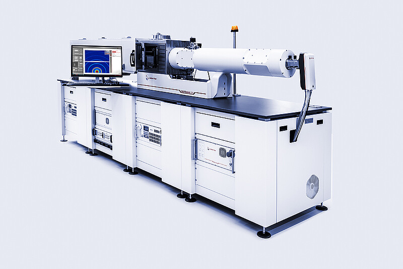
- SAXS/WAXS/GISAXS/RheoSAXS laboratory beamline: SAXSpoint 5.0
Source:
Hassett, Kimberly J., et al. “Impact of Lipid Nanoparticle Size on MRNA Vaccine Immunogenicity.” Journal of Controlled Release, vol. 335, July 2021, pp. 237–46.
Patel, Siddharth, et al. “Naturally-Occurring Cholesterol Analogues in Lipid Nanoparticles Induce Polymorphic Shape and Enhance Intracellular Delivery of MRNA.” Nature Communications, vol. 11, no. 1, Dec. 2020, p. 983.