Liposomes and Their Applications
Introduction
Liposomes are artificial vesicles with a double lipid layer. They represent one of the most promising carriers not only for biologically active compounds in cosmetic and pharmaceutical applications, but also for substances used in the food industry (e.g., antioxidants, flavors, antimicrobials) that are incorporated into liposomes in order to make them stable.
The liposomes’ particle size, as well as surface charge, affect the encapsulation capacity and the uptake from the target cells, especially if they are used as drug delivery systems. Any modification of the liposome surface can also be monitored via measurement of the zeta potential.
Knowledge of the liposome size as well as liposome zeta potential helps in the characterization of the special coatings and in the controlling of aggregation, fusion, and precipitation, which are important factors affecting the stability of liposomal formulations and the in-vivo behavior of liposomes.
What are liposomes made of?
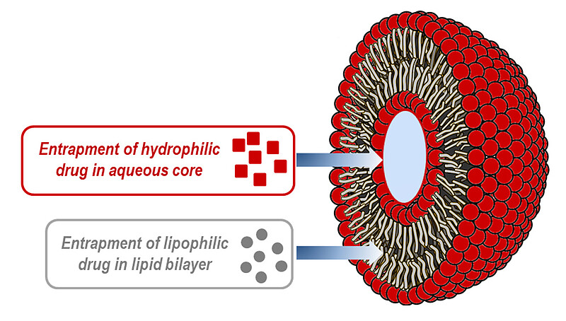
Liposomes consist of phospholipids (e.g., lecithin) which are amphiphilic molecules that have a hydrophilic head and a lipophilic tail. When these phospholipids are introduced into an aqueous medium, they self-assemble into vesicles with the polar ends facing the aqueous medium and non-polar ends forming a bilayer – as shown in Figure 1. Due to this unique property, they can contain a wide variety of hydrophilic (in the hydrophilic core) and hydrophobic components (in the hydrophobic double layer).
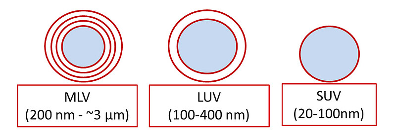
Liposomes and nanoliposomes (liposomes in the nanoscale) can be formed by a combination of triglycerides or mono/diglycerides and phospholipids. In addition to lipid and/or phospholipid molecules, nanoliposomes may contain other molecules, such as sterols, in their structure. The most widely used sterol in the manufacture of the lipid vesicles is cholesterol, which helps increase the stability of the vesicles by reducing the permeability of the lipid membrane to solutes [1]. According to the size and the number of layers, three main categories of liposomes can be identified (Figure 2) [2]:
- Small unilamellar vesicles (SUVs): single phospholipid bilayer, 20 nm to 100 nm
- Large unilamellar vesicles (LUVs): single phospholipid bilayer, 100 nm to 400 nm
- Multilamellar large vesicles (MLVs): multiple phospholipid bilayer, 500 nm to ~3000 nm
All three categories are mostly used for drug delivery. The main difference is the clearance from the bloodstream by the immune system. The large multilamellar vesicle size causes a faster removal due to the interaction with blood proteins.
Liposomes can be coated by molecules such as PEG, silica, chitosan, and polysaccharide, which build a hydrophilic and steric barrier. The modification is important when liposomes are used for drug delivery systems. The change of the surface charge and the hindrance increase prevent the binding of plasma proteins and their recognition by some cellular receptors, which might cause the removal from the circulation system. By means of the coating, the stability of liposomes increases, and the gradual release of the incorporated drug can be ensured. [3]
Importance of liposome particle size
The characterization of liposome size is important not only for checking the stability but also for the final application. In the field of drug-delivery systems, liposome diameter may affect cell interaction and subsequent uptake as well as the amount of encapsulated drug. For these reasons, the liposome size should be monitored during the formulation phase in order to ensure a product has the desired properties. After generation of liposomes with the required size, further tests can be performed to investigate stability at different conditions (e.g., temperature, pH, ionic strength).
Monitoring the formulation process
Liposome formulation can be performed using different methods according to the desired liposome diameter. One of the most common methods consists of the dispersion of lipids in an organic solvent, evaporation of the solvent, and resuspension of dry liposomes in aqueous systems.
In pharmaceutical formulations (e.g., vaccines), the smallest liposomes, such as SUVs, are often used because their small size reduces clearance from the systemic circulation. Therefore, they circulate longer and have an increased probability of exerting the desired therapeutic effect in tissues – although the encapsulation capacity is limited because of the liposome size.
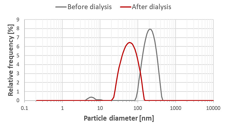
One of the formulation methods of SUVs consists of mixing surfactants and lipids in the organic solvent (e.g., dichloromethane). After removal of surfactants, lipids self-assemble to form the liposomes. The process of surfactant removal and liposome formation can be monitored using both particle size and zeta potential. Figure 3 shows particle size results obtained before and after surfactant removal by means of dialysis. The shift of the size from 200 nm towards 60 nm indicates the formation of SUVs.
Monitoring the stability
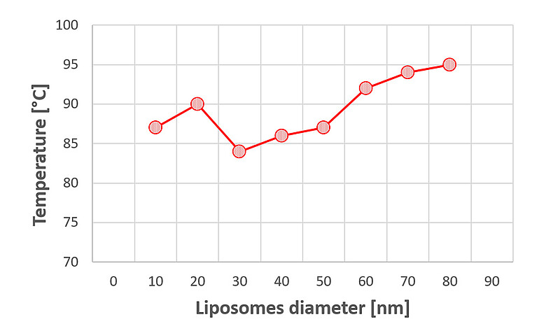
Some of the factors that can affect the liposomes’ stability are pH, ionic strength, and temperature. At elevated temperatures lower than their final melting point (Tm), they undergo a phase transition that alters their permeability. At the gel-to-liquid-crystalline-transition temperature (Tc), the lipidic bilayer loses its ordered packing, and its fluidity increases [1]. At the same time, liposome fusion starts and aggregation occurs [4]. Figure 4 shows how increasing the temperature causes a consistent increase in the liposome size.
The increase of pH contributes to a reduction in the liposomes’ shelf-life because the phospholipids get protonated and build hydrogen bonds with the neighboring phospholipid molecules [4]. Furthermore, the increase of the ionic strength of the medium can cause compression of the electrical double layer and reduce the electrostatic repulsion between liposome particles [5].
Importance of liposome zeta potential
As soon as liposomes form, they develop a surface charge at the interface in contact with the surrounding media, and the zeta potential can be measured. The liposome zeta potential provides not only information about the surface properties (e.g., cationic, anionic) but also about the stability as well as the destiny in a living organism. In fact, liposomes used as drug delivery systems interact with protein membranes and permeate into the cell only if they present the appropriate charge.
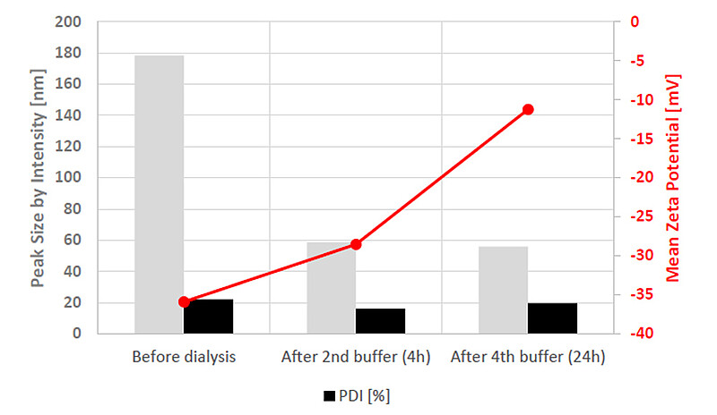
Figure 5 displays how the liposome zeta potential and particle size change during the progressive removal of surfactant. At the end of the process, the measured liposomes’ diameter is 60 nm, and the charge is slightly negative. In fact, the surfactant introduces negative charges on the vesicles. By removing the surfactants, fewer negative charges will be at the vesicles/water interface, and this determines a decrease in the zeta potential magnitude.
The information about the liposome zeta potential is important for nanoparticles with drug delivery function because it determines the interaction with the cells. In fact, the negative charge contributes to suppression of liposome uptake from cells (e.g., macrophages), which are involved in the defense against infectious agents [6].
References
- Mozafari, Marcel R. 2010. “Nanoliposomes: Preparation and Analysis.” Methods in Molecular Biology 605: 29–50. doi.org/10.1007/978-1-60327-360-2_2.
- Yang, Feng, Chen Jin, Yongjian Jiang, Ji Li, Yang Di, Quanxing Ni, and Deliang Fu. 2011. “Liposome Based Delivery Systems in Pancreatic Cancer Treatment: From Bench to Bedside.” Cancer Treatment Reviews 37 (8): 633–642. doi.org/10.1016/j.ctrv.2011.01.006.
- Sagristá, Maria, Margarita Mora, and M. Africa De Madariaga. 2000. “Surface Modified Liposomes by Coating with Charged Hydrophilic Molecules.” Cellular and Molecular Biology Letters 5: 19–33.
- Moh’d Atrouse, Omar. 2002. “The Effects of Liposome Composition and Temperature on the Stability of Liposomes and the Interaction of Liposomes with Human Neutrophils.” Pakistan Journal of Biological Sciences 5: 948–951. doi.org/10.3923/pjbs.2002.948.951.
- Wang, Xuejiao, Caleb John Swing, Tingting Feng, Shuqin Xia, Jingyang Yu, and Xiaoming Zhang. 2020. “Effects of Environmental pH and Ionic Strength on the Physical Stability of Cinnamaldehyde-loaded Liposomes.” Journal of Dispersion Science and Technology 41 (10): 1568–1575. doi.org/10.1080/01932691.2019.1627887.
- Xuedong, Yan, Gerrit L. Scherphof, and Jan A. Kamps. 2005. “Liposome Opsonization.” Journal of Liposome Research 15 (1–2): 109–39. doi.org/10.1081/lpr-64971.